Lesson Objectives
- State the cell theory, and list the discoveries that led to it.
- Describe the diversity of cell shapes, and explain why cells are so small.
- Identify the parts that all cells have in common.
- Contrast prokaryotic and eukaryotic cells.
WORKBOOK ASSIGNMENT
Chapter 3.1 workbook pages
Get the workbook here: https://guesthollow.com/store/free-high-school-biology-workbook/
Vocabulary
- cytoplasm
- all of the material inside the plasma membrane of a cell (excluding organelles)
- eukaryote
- organism that has cells containing a nucleus and other organelles
- eukaryotic cell
- cell that contains a nucleus and other organelles
- nucleus (plural, nuclei)
- organelle inside eukaryotic cells that contains most of the cell’s DNA and acts as the control center of the cell
- organelle
- structure within the cytoplasm of a cell that is enclosed within a membrane and performs a specific job
- plasma membrane
- thin coat of lipids (phospholipids) that surrounds and encloses a cell
- prokaryote
- single-celled organism that lacks a nucleus
- prokaryotic cell
- cell without a nucleus that is found in single-celled organisms
- ribosome
- organelle inside all cells where proteins are made
- virus
- tiny, nonliving particle that contains DNA but lacks other characteristics of living cells
Introduction
If you look at living matter with a microscope—even a simple light microscope—you will see that it consists of cells. Cells are the basic units of the structure and function of living things. They are the smallest units that can carry out the processes of life. All organisms are made up of one or more cells, and all cells have many of the same structures and carry out the same basic life processes. Knowing the structures of cells and the processes they carry out is necessary to understanding life itself.
Discovery of Cells
The first time the word cell was used to refer to these tiny units of life was in 1665 by a British scientist named Robert Hooke. Hooke was one of the earliest scientists to study living things under a microscope. The microscopes of his day were not very strong, but Hooke was still able to make an important discovery. When he looked at a thin slice of cork under his microscope, he was surprised to see what looked like a honeycomb. Hooke made the drawing in Figure below to show what he saw. As you can see, the cork was made up of many tiny units, which Hooke called cells.

Leeuwenhoek’s Discoveries
Soon after Robert Hooke discovered cells in cork, Anton van Leeuwenhoek (lee-oon-hook) in Holland made other important discoveries using a microscope. Leeuwenhoek made his own microscope lenses, and he was so good at it that his microscope was more powerful than other microscopes of his day. In fact, Leeuwenhoek’s microscope was almost as strong as modern light microscopes. Using his microscope, Leeuwenhoek discovered tiny animals such as rotifers. The magnified image of a rotifer in Figure below is similar to what Leeuwenhoek observed. Leeuwenhoek also discovered human blood cells. He even scraped plaque from his own teeth and observed it under the microscope. What do you think Leeuwenhoek saw in the plaque? He saw tiny living things with a single cell that he named animalcules (“tiny animals”). Today, we call Leeuwenhoek’s animalcules bacteria.

The Cell Theory
By the early 1800s, scientists had observed the cells of many different organisms. These observations led two German scientists, named Theodor Schwann and Matthias Jakob Schleiden, to propose that cells are the basic building blocks of all living things. Around 1850, a German doctor named Rudolf Virchow was studying cells under a microscope when he happened to see them dividing and forming new cells. He realized that living cells produce new cells through division. Based on this realization, Virchow proposed that living cells arise only from other living cells. The ideas of all three scientists—Schwann, Schleiden, and Virchow—led to the cell theory, which is one of the fundamental theories of biology. The cell theory states that:
- All organisms are made of one or more cells.
- All the life functions of organisms occur within cells.
- All cells come from already existing cells.
Microscopes
Starting with Robert Hooke in the 1600s, the microscope opened up an amazing new world—the world of life at the level of the cell. As microscopes continued to improve, more discoveries were made about the cells of living things. However, by the late 1800s, light microscopes had reached their limit. Objects much smaller than cells, including the structures inside cells, were too small to be seen with even the strongest light microscope. Then, in the 1950s, a new type of microscope was invented. Called the electron microscope, it used a beam of electrons instead of light to observe extremely small objects. With an electron microscope, scientists could finally see the tiny structures inside cells. In fact, they could even see individual molecules and atoms. The electron microscope had a huge impact on biology. It allowed scientists to study organisms at the level of their molecules and led to the emergence of the field of molecular biology. With the electron microscope, many more cell discoveries were made. Figure below shows how the cell structures called organelles appear when scanned by an electron microscope.

KQED: The World’s Most Powerful Microscope
Lawrence Berkeley National labs uses a $27 million electron microscope to make images to a resolution of half the width of a hydrogen atom. This makes it the world’s most powerful microscope.
KQED The World’s Most Power Microscope:
KQED: Confocal Microscopy
Cutting-edge microscopes, called confocal microscopes, at the University of California, San Francisco are helping scientists create three-dimensional images of cells, and may help lead to new medical breakthroughs, including a treatment for Type 1 diabetes.
Diversity of Cells
Today, we know that all living cells have certain things in common. For example, all cells share functions such as obtaining and using energy, responding to the environment, and reproducing. We also know that different types of cells—even within the same organism—may have their own unique functions as well. Cells with different functions generally have different shapes that suit them for their particular job. Cells vary in size as well as shape, but all cells are very small. In fact, most cells are much smaller than the period at the end of this sentence. If cells have such an important role in living organisms, why are they so small? Even the largest organisms have microscopic cells. What limits cell size?
Cell Size
The answer to these questions is clear once you know how a cell functions. To carry out life processes, a cell must be able to quickly pass substances into and out of the cell. For example, it must be able to pass nutrients and oxygen into the cell and waste products out of the cell. Anything that enters or leaves a cell must cross its outer surface. It is this need to pass substances across the surface that limits how large a cell can be. Look at the two cubes in Figure below. As this figure shows, a larger cube has less surface area relative to its volume than a smaller cube. This relationship also applies to cells; a larger cell has less surface area relative to its volume than a smaller cell. A cell with a larger volume also needs more nutrients and oxygen and produces more wastes. Because all of these substances must pass through the surface of the cell, a cell with a large volume will not have enough surface area to allow it to meet its needs. The larger the cell is, the smaller its ratio of surface area to volume, and the harder it will be for the cell to get rid of its wastes and take in necessary substances. This is what limits the size of the cell.

Why are Cells Small?
Cell Shape
Cells with different functions often have different shapes. The cells pictured inFigure below are just a few examples of the many different shapes that cells may have. Each type of cell in the figure has a shape that helps it do its job. For example, the job of the nerve cell is to carry messages to other cells. The nerve cell has many long extensions that reach out in all directions, allowing it to pass messages to many other cells at once. Do you see the tail-like projections on the algae cells? Algae live in water, and their tails help them swim. Pollen grains have spikes that help them stick to insects such as bees. How do you think the spikes help the pollen grains do their job? (Hint: Insects pollinate flowers.)

As these pictures show, cells come in many different shapes. Clockwise from the upper left photo are a nerve cell, red blood cells, bacteria, pollen grains, and algae. How are the shapes of these cells related to their functions?
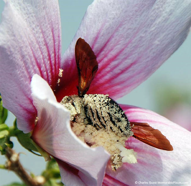
Notice the pollen grains sticking to the bee’s body. The grains are able to stick due to the spikes they are covered with. How could pollen evolve spikes? If they didn’t have spikes on them in the first place, they wouldn’t stick to insects. If they didn’t stick to insects, they wouldn’t be able to travel to other flowers for the purposes of pollination. Plants that are currently pollinated by insects would never have become pollinated which means they wouldn’t have survived. God created pollen cells to have spikes just for this purpose. (Photo by Charles Guest of Memorable Places Photography)
Parts of a Cell
Although cells are diverse, all cells have certain parts in common. The parts include a plasma membrane, cytoplasm, ribosomes, and DNA.
- The plasma membrane (also called the cell membrane) is a thin coat of lipids that surrounds a cell. It forms the physical boundary between the cell and its environment, so you can think of it as the “skin” of the cell.
- Cytoplasm refers to all of the cellular material inside the plasma membrane. Cytoplasm is made up of a watery substance called cytosol and contains other cell structures such as ribosomes.
- Ribosomes (righ-boe-somes) are structures in the cytoplasm where proteins are made.
- DNA is a nucleic acid found in cells. It contains the genetic instructions that cells need to make proteins.

These parts are common to all cells, from organisms as different as bacteria and human beings. How did all known organisms come to have such similar cells? The similarities show that God used a similar blueprint when creating life.
A nice introduction to the cell is available at http://www.youtube.com/watch?v=Hmwvj9X4GNY (21:03). This video is OPTIONAL.
There is another basic cell structure that is present in many but not all living cells: the nucleus. The nucleus of a cell is a structure in the cytoplasm that is surrounded by a membrane (the nuclear membrane) and contains DNA. Based on whether they have a nucleus, there are two basic types of cells: prokaryotic cells and eukaryotic cells.
Prokaryotic Cells
Prokaryotic (proe-care-ee-ot-ick) cells are cells without a nucleus. The DNA in prokaryotic cells is in the cytoplasm rather than enclosed within a nuclear membrane. Prokaryotic cells are found in single-celled organisms, such as bacteria, like the one shown in Figure below. Organisms with prokaryotic cells are called prokaryotes.

Bacteria are described in the following video http://www.youtube.com/watch?v=TDoGrbpJJ14 (18:26). This video is OPTIONAL.
Eukaryotic Cells
Eukaryotic (Yoo-care-ee-ot-ick) cells are cells that contain a nucleus. A typical eukaryotic cell is shown in Figure below. Eukaryotic cells are usually larger than prokaryotic cells, and they are found mainly in multicellular organisms. Organisms with eukaryotic cells are called eukaryotes, and they range from fungi to people. Eukaryotic cells also contain other organelles besides the nucleus. An organelle is a structure within the cytoplasm that performs a specific job in the cell. Organelles called mitochondria, for example, provide energy to the cell, and organelles called vacuoles store substances in the cell. Organelles allow eukaryotic cells to carry out more functions than prokaryotic cells can.

Viruses: Prokaryotes or Eukaryotes?
Viruses, like the one depicted in Figure below, are tiny particles that may cause disease. Human diseases caused by viruses include the common cold and flu. Do you think viruses are prokaryotes or eukaryotes? The answer may surprise you. Viruses are not cells at all, so they are neither prokaryotes nor eukaryotes.

Viruses contain DNA but not much else. They lack the other parts shared by all cells, including a plasma membrane, cytoplasm, and ribosomes. Therefore, viruses are not cells, but are they alive? All living things not only have cells; they are also capable of reproduction. Viruses cannot reproduce by themselves. Instead, they infect living hosts, and use the hosts’ cells to make copies of their own DNA. For these reasons, most scientists do not consider viruses to be living things.
An overview of viruses can be seen at http://www.youtube.com/watch?v=0h5Jd7sgQWY (23:17). This video is OPTIONAL.
Lesson Summary
- Discoveries about cells using the microscope led to the development of the cell theory. This theory states that all organisms are made of one or more cells, all the life functions of organisms occur within cells, and all cells come from already existing cells.
- All cells are very small because they need to pass substances across their surface. Their small size gives them a relatively large ratio of surface area to volume, facilitating the transfer of substances. The shapes of cells may vary, and a cell’s shape generally suits its function.
- Cells are diverse, but all cells contain a plasma membrane, cytoplasm, ribosomes, and DNA.
- Prokaryotic cells are cells without a nucleus. They are found in single-celled organisms. Eukaryotic cells are cells with a nucleus and other organelles. They are found mainly in multicellular organisms.
Review Questions
Recall
1. What did Hooke and Leeuwenhoek discover about cells by using a microscope?
2. What does the cell theory state? Name the three scientists mainly responsible for developing the cell theory.
3. List the four parts that are found in all living cells.
Apply Concepts
4. One of the cells pictured below is a human brain cell. The other cell is found in human blood. Which cell came from the brain? Explain your answer.

Think Critically
5. Why are all cells very small? Explain what limits the size of cells.
6. Compare and contrast prokaryotic cells and eukaryotic cells.
7. Explain why viruses are not considered to be living.
Points to Consider
Cells have many different structures that carry out the processes of life.
- Beside the cell parts described in this lesson, what other structures do you think cells might have? What life processes might these other structures carry out?
- Do you think plant and animal cells are just alike? Or do they differ in some way? How might they differ?
Previous: Water, Acids, Bases
Next: Cell Structures

CK-12 Foundation is licensed under Creative Commons AttributionNonCommercial 3.0 Unported (CC BY-NC 3.0)”
• Terms of Use • Attribution


Hey, just wanted to let you guys know that the KQED links did not work for me. This could be a problem on my end, but I just wanted to let you guys know.
Thank you SO much for letting us know! I’ll check them out asap, and if there is an issue on my end too, I’ll take them down. 🙂 We so appreciate you! Please don’t hesitate to let us know if you run into any other issues!